


陶可医生的科普号
- 视频 髋关节核心肌群锻炼小视频
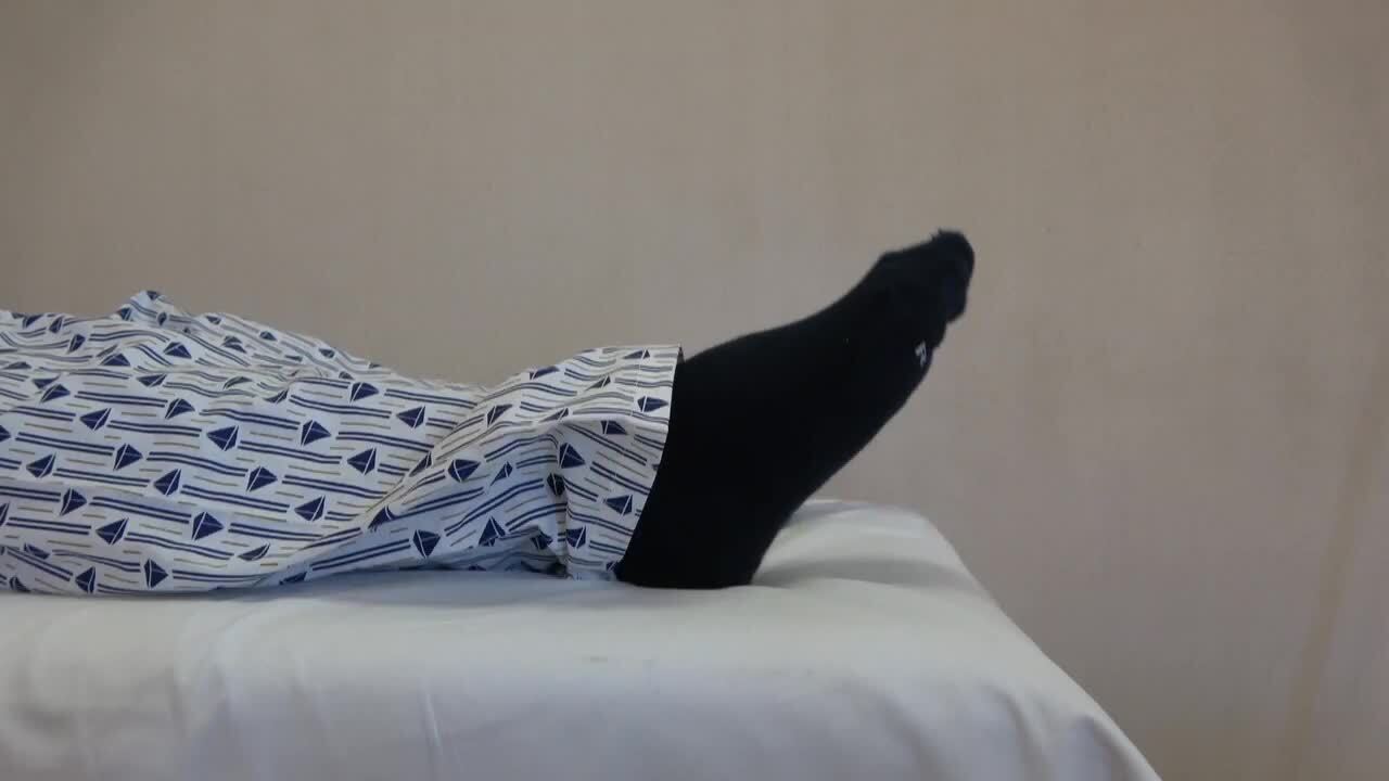
PAO截骨术后1双脚踝泵训练2股四头肌锻炼-平卧位3股四头肌锻炼-坐位4蚌式运动5侧卧位-侧抬大腿6侧卧位-侧抬大腿-2,加强锻炼(5公斤弹力带)7大腿绷紧后方抬起锻炼8单腿后伸9单腿后伸-加强锻炼(5公斤弹力带)10单屈髋-双屈髋11仰卧位-足踝部滚动锻炼12非负重蹲起锻炼13站立位,大腿伸直向侧方抬起锻炼(外展肌群:臀中肌、臀小肌、阔筋膜张肌)14大腿侧后伸直抬起锻炼(后伸肌群:臀大肌)15双下肢前后交叉锻炼(内收肌群:长收肌、大收肌、耻骨肌等)16核心肌群训练模块-1:靠墙蹲起锻炼16核心肌群训练模块-2:靠墙蹲起锻炼17核心肌群训练模块-1——太极式蹲起锻炼18改良平板支撑训练19核心肌群训练模块-3——站立伸腿、屈髋屈膝20核心肌群训练模块-4——弓步箭步蹲式训练
 陶可 主治医师 北京大学人民医院 骨关节科2917人已读
 - 学术前沿 2018年美国华盛顿大学:分类简述:股骨头骨折的Pipkin分类
2018年美国华盛顿大学:分类简述:股骨头骨折的Pipkin分类译者:陶可(北京大学人民医院骨关节科) 文献出处:Nicholas M Romeo, Reza Firoozabadi. Classif
陶可 主治医师 北京大学人民医院 骨关节科2279人已读 - 学术前沿 2014年意大利米兰大学与英国利兹大学:使用(骨)诱导小室技术和基于生物学方法治疗股骨头坏死:适应症
2014年意大利米兰大学与英国利兹大学:使用(骨)诱导小室技术和基于生物学方法治疗股骨头坏死:适应症和临床结果译者:陶可(北京大学人民医院骨关节科)摘要:目的:确定髓心减压(CD)技术联合重组骨形态发生蛋白、自体间充质干细胞(MSCs)和异体骨替代物进入股骨头坏死病灶对临床症状和股骨头坏死进展的疗效。患者和方法:对 38 名早期股骨头坏死患者(40 髋)进行了为期 4 年的研究。结果:CD 技术结合重组形态发生蛋白、自体 MSCs 和异体骨移植替代物与疼痛和关节症状的显着减少相关,并降低了骨折分期的发生率。在 36 个月时,33 名患者实现了临床和放射学愈合。结论:本长期随访研究证实,CD技术联合重组骨形态发生蛋白、自体MSCs和异体移植骨替代物可能是治疗早期股骨头坏死患者的有效方法。关键词:股骨头坏死AVN;自体植入;缺血性坏死;生物室;髓心减压;早期;股骨头;间充质干细胞;骨坏死;重组骨形态发生蛋白rhBMP。文献出处:G M Calori, E Mazza, M Colombo, S Mazzola, G V Mineo, P V Giannoudis. Treatment of AVN using the induction chamber technique and a biological-based approach: indications and clinical results. Injury. 2014 Feb;45(2):369-73.doi: 10.1016/j.injury.2013.09.014. Epub 2013 Sep 19.Treatment of AVN using the induction chamber technique and a biological-based approach: indications and clinical results.AbstractObjective: To determine the efficacy of core decompression (CD) technique combined with recombinant morphogenetic proteins, autologous mesenchymal stem cells (MSCs) and xenograft bone substitute into the necrotic lesion of the femoral head on clinical symptoms and on the progression of osteonecrosis of the femoral head.Patients and methods: A total of 38 patients (40 hips) with early stage osteonecrosis of the femoral head were studied over a 4-year period.Results: CD technique combined with recombinant morphogenetic proteins, autologous MSCs and xenograft bone substitute was associated with a significant reduction in both pain and joint symptoms and reduced the incidence of fractural stages. At 36 months, 33 patients achieved clinical and radiographic healing.Conclusion: This long-term follow-up study confirmed that CD technique combined with recombinant morphogenetic proteins, autologous MSCs and xenograft bone substitute may be an effective treatment for patients with early stage osteonecrosis of the femoral head.Keywords: AVN; Autologous implantation; Avascular necrosis; Biological chamber; Core decompression; Early stage; Femoral head; Mesenchymal stem cells; Osteonecrosis; rhBMP.Fig. 1. Two cases of AVN of the femoral head treated with our technique. (a–c) First patient, 33-year-old man. (a) MRI image showing the lesion; (b) MRI image of the femoral head 1 year after the procedure showing the implanted biotechnologies and the conserved shape of the femoral head; (c) CT scan after 3 years, good ossification of the subcondral bone, no trabecular collapse, shape of the femoral head still conserved. (d–f) second patient, 54-year-old woman. (d) MRI image showing the lesion; (e) X-ray at 2 years after the treatment, good integration of the biotechnologies implanted, no collapse; (f) CT scan after 3 years, no collapse, shape of the head conserved.图 1. 使用我们的技术治疗的两例股骨头 AVN。 (a-c) 第一位患者,33 岁男性。 (a) 显示病变的 MRI 图像; (b) 手术后 1 年的股骨头 MRI 图像,显示植入的生物技术和股骨头的保留形状; (c) 3年后CT扫描,软骨下骨骨化良好,无小梁塌陷,股骨头形态尚存。 (d-f) 第二位患者,54 岁女性。 (d) 显示病变的 MRI 图像; (e) 治疗后2年X线片,植入的生物技术整合良好,无塌陷; (f) 3年后CT扫描,无塌陷,股骨头形状保存。注:(骨)诱导小室技术Giorgio M Calori, Peter V Giannoudis. Enhancement of fracture healing with the diamond concept: the role of the biological chamber. Injury. 2011 Nov;42(11):1191-3.doi: 10.1016/j.injury.2011.04.016. Epub 2011 May 18.Fig. 1. Diamond concept and the biological chamber. V = vascularity, H = host, MS = mechanical stability, MSC = osteoprogenitor cells, S = scaffold, GF = growth factor. 1. Closed chamber; 2. Open chamber; 3. Partially closed chamber.图 1. 菱形概念和生物小室。 V = 血管分布,H = 宿主,MS = 机械稳定性,MSC = 骨祖细胞,S = 生物支架,GF = 生长因子。 1.密闭室; 2.开放室; 3. 部分封闭的腔室。
陶可 主治医师 北京大学人民医院 骨关节科770人已读 - 学术前沿 2018年日本筑波大学医学院:系统性红斑狼疮皮质类固醇相关股骨头坏死的自体骨髓穿刺浓缩移植中期结果
系统性红斑狼疮皮质类固醇(治疗)相关股骨头坏死的自体骨髓穿刺浓缩移植中期结果译者:陶可(北京大学人民医院骨关节科)摘要:目的:我们之前已经建立了浓缩自体骨髓穿刺移植(CABMAT),这是一种用于治疗股骨头坏死(ONFH)的一步式、微创、保留髋关节的手术技术。本研究旨在评估 CABMAT 作为保留髋关节手术方法的效果,防止系统性红斑狼疮 (SLE) 患者转为全髋关节置换术 (THA) 和股骨头塌陷。方法:自2003年以来,52例SLE患者(男8例,女44例,92髋,平均年龄35.3(16-77)(岁))接受CABMAT治疗,平均随访5.5(0.7-14)年。对THA的发生率及其预测因素进行了分析。结果:THA 的总体转化率为 29% (27/92)。在 A、B、C1 和 C2 型中,THA 的转化率分别为 0% (0/3)、0% (0/4)、22% (9/41) 和 41% (18/44)。在第 1、2、3A、3B 和4阶段,向 THA 的转化率分别为 26% (5/19)、26% (6/23)、28% (11/39)、44% (4/9) 和 50% (1/2)。在多元逻辑回归分析中,性别、体重指数(BMI)、术前ONFH类型和术前分期与转换为 THA 显著相关。结论:THA的转化率低于自然疗程髓心减压术,但高于其他骨髓移植和截骨术。由于性别、术前疾病类型和术前分期与转换为 THA 显著相关,因此推测女性、晚期(3A 期或以上)和晚期(C 型或以上)比例较高影响了这项研究的THA 转化率。关键词:浓缩自体骨髓穿刺移植;生长因子;髋关节保留手术;间充质干细胞;股骨头坏死;系统性红斑狼疮文献出处:Yohei Tomaru, Tomokazu Yoshioka, Hisashi Sugaya, Yukiyo Shimizu, Katsuya Aoto, Hiroshi Wada, Hiroshi Akaogi, Masashi Yamazaki, Hajime Mishima. Mid-term results of concentrated autologous bone marrow aspirate transplantation for corticosteroid-associated osteonecrosis of the femoral head in systemic lupus erythematosus. Int Orthop. 2018 Jul;42(7):1623-1630.doi: 10.1007/s00264-018-3959-y. Epub 2018 Apr 28.Mid-term results of concentrated autologous bone marrow aspirate transplantation for corticosteroid-associated osteonecrosis of the femoral head in systemic lupus erythematosus.AbstractPurpose: We had previously established concentrated autologous bone marrow aspirate transplantation (CABMAT), a one-step, low-invasive, joint-preserving surgical technique for treating osteonecrosis of the femoral head (ONFH). This study aimed to evaluate the effects of CABMAT as a hip-preserving surgical approach, preventing conversion to total hip arthroplasty (THA) and femoral head collapse in patients with systemic lupus erythematosus (SLE).Methods: Since 2003, 52 SLE patients (8 male, 44 female, 92 hips, mean age 35.3 (16-77) (years) were treated with CABMAT. The mean follow-up period was 5.5 (0.7-14) years. Conversion rate to THA and its predicting factors were analyzed.Results: The overall conversion rate to THA was 29% (27/92). Conversion rate to THA was 0% (0/3), 0% (0/4), 22% (9/41), and 41% (18/44) in types A, B, C1, and C2, respectively. Conversion rate to THA was 26% (5/19), 26% (6/23), 28% (11/39), 44% (4/9), and 50% (1/2) in stages 1, 2, 3A, 3B, and 4, respectively. In multivariate logistic regression analysis, sex, body mass index (BMI), pre-operative type, and pre-operative stage were significantly correlated with conversion to THA.Conclusion: The conversion rate to THA was lower than that in the natural course and core decompression, but was higher than that seen in other bone marrow transplantation and osteotomy. Since sex, pre-operative type, and pre-operative stage were significantly correlated with conversion to THA, it is suggested that the higher proportion of women, advanced stage (stage 3A or above), and advanced type (type C or above) in this study affected the THA conversion rate.Keywords: Concentrated autologous bone marrow aspirate transplantation; Growth factors; Joint preserving surgery; Mesenchymal stem cells; Osteonecrosis of the femoral head; Systemic lupus erythematosus.Fig. 1 A 58-year-old woman, SLE, no collapse progression after 5 years. Pre-operative plane radiograph: AP view (a), frog leg lateral view (b). Five years after operation: AP view (c), frog leg lateral view (d). Preoperative MRI (T1WI): coronal view (e), oblique axial view (f). Five years after operation: coronal view (g), oblique axial view (h).图 1 一名 58 岁女性,SLE,5 年后无塌陷进展。术前平片:前后AP位 视图 (a),蛙式位视图 (b)。术后五年:前后AP位视图 (c),蛙式位视图 (d)。术前 MRI (T1WI):冠状位视图 (e),斜轴位视图 (f)。术后五年:冠状位(g),斜轴位(h)。Fig. 2 Prognosis of stage 1 and 2 hips. (a) THA conversion rate to THA in stage 1 and 2. (b) Stage at most recent follow-up in the THA nonconversion group. Definition of stage is as follows. Stage 1: No abnormality is detected on radiographs. Abnormality can be detected only on MRI or scintigraphy. Stage 2: Sclerotic change without collapse is detected on radiographs. Stage 3A: collapse of femoral head less than 3 mm, without osteoarthritic change. Stage 3B: collapse of femoral head more than 3 mm, without osteoarthritic change. Stage 4: Osteoarthritic change is detected. MRI magnetic resonance imaging, THA total hip arthroplasty.图 2 1 期和 2 期髋关节的预后。 (a) 第 1 阶段和第 2 阶段的 THA 转换率。 (b) THA 未转换组最近一次随访时的阶段。阶段的定义如下。第一阶段:X光片未发现异常。异常只能在 MRI 或闪烁扫描中检测到。阶段 2:在 X 光片上检测到没有塌陷的硬化变化。 3A 期:股骨头塌陷小于 3 毫米,无骨关节炎改变。 3B 期:股骨头塌陷超过 3 毫米,无骨关节炎改变。第 4 阶段:检测到骨关节炎变化。 MRI 磁共振成像,THA 全髋关节置换术。
陶可 主治医师 北京大学人民医院 骨关节科858人已读 - 学术前沿 2019年日本筑波大学医学院:浓缩自体骨髓穿刺移植治疗股骨头坏死的10年(随访)结果:一项回顾性研究
浓缩自体骨髓穿刺移植治疗股骨头坏死的10年(随访)结果:一项回顾性研究译者:陶可(北京大学人民医院骨关节科)摘要:背景:特发性股骨头坏死 (ONFH) 发生在相对年轻的患者。因此,在这些患者中防止由此产生的股骨头塌陷和全髋关节置换术是很重要的。2003 年,我们在筑波大学医学院启动了自体骨髓穿刺浓缩移植 (CABMAT),这是一种保留髋关节的 ONFH 治疗方法。在这里,我们报告了 CABMAT 治疗的长期结果。方法:我们回顾性整理和分析了 2003 年 4 月至 2008 年 4 月间接受 CABMAT 治疗的 69 例特发性 ONFH 患者(109 髋)的人口统计学和治疗数据。结果:共44例(男21例,女23例,80髋)完成10年随访。随访率为 73.4%,平均随访时间为 12.0(范围,10.0-15.4)年。患者的平均年龄为 42.2(范围,16.3-70.5)岁。使用骨循环研究协会(ARCO) 分类系统进行术前分析,分别将 12、31、32 和 5 个髋关节分为 1、2、3 和 4 期。全髋关节置换术 (THA) 的总转换率为 34%(27/80 髋)。在多元回归分析中,发现 ONFH 的术前分期和体重指数与转换为 THA 显著相关。总共有 43 个髋关节(共 80 个)被归类为塌陷前阶段(即第 1 或第 2 阶段)。这些髋关节的整体塌陷率和 THA 转换率估计分别为 49% (21/43) 和 14% (6/43)。结论:根据我们的长期研究结果,微创可行的CABMAT治疗可作为ONFH的一种保髋治疗方法。关键词:骨髓穿刺浓缩液;保髋手术;股骨头坏死文献出处:Yohei Tomaru, Tomokazu Yoshioka, Hisashi Sugaya, Hiroshi Kumagai, Kojiro Hyodo, Katsuya Aoto, Hiroshi Wada, Hiroshi Akaogi, Masashi Yamazaki, Hajime Mishima. Ten-year results of concentrated autologous bone marrow aspirate transplantation for osteonecrosis of the femoral head: a retrospective study. BMC Musculoskelet Disord. 2019 Sep 5;20(1):410.doi: 10.1186/s12891-019-2797-4.Ten-year results of concentrated autologous bone marrow aspirate transplantation for osteonecrosis of the femoral head: a retrospective study.AbstractBackground: Idiopathic osteonecrosis of the femoral head (ONFH) occurs at a relatively younger age. It is therefore important to prevent the resultant femoral head collapse and requirement of total hip arthroplasty in these patients. In 2003, we initiated concentrated autologous bone marrow aspirate transplantation (CABMAT), a joint-preserving treatment for ONFH, at our institution. Here, we report the long-term results of CABMAT treatment.Methods: We retrospectively collated and analyzed the demographic and treatment data of 69 patients (109 hips) with idiopathic ONFH treated with CABMAT between April 2003 and April 2008.Results: Totally, 44 patients (21 men, 23 women, 80 hips) completed the 10-year follow-up. The follow-up rate was 73.4%, and the mean follow-up period was 12.0 (range, 10.0-15.4) years. The mean age of the patients was 42.2 (range, 16.3-70.5) years. Using the Association Research Circulation Osseous (ARCO) classification system for preoperative analysis, 12, 31, 32, and 5 hips were classified as stages 1, 2, 3, and 4, respectively. The overall rate of conversion to total hip arthroplasty (THA) was 34% (27/80 hips). In a multivariate regression analysis, the preoperative stage of ONFH and the body mass index were found to correlate significantly with conversion to THA. Totally, 43 hips (of 80) were classified as belonging to the pre-collapse stage (i.e., stages 1 or 2). The overall collapse rate and the THA-conversion rate of these hips were estimated to be 49% (21/43) and 14% (6/43), respectively.Conclusions: On the basis of our long-term findings, the minimally invasive and feasible CABMAT therapy can be utilized as one of a joint-preserving treatment for ONFH.Keywords: Bone marrow aspirate concentrate; Hip preserving surgery; Osteonecrosis of the femoral head.Fig. 1 Survival curve (end point: conversion to total hip arthroplasty).图 1 生存曲线(终点:转换为全髋关节置换术)。
陶可 主治医师 北京大学人民医院 骨关节科1126人已读 - 学术前沿 2019年最新:股骨头坏死:病理生理学和当前治疗概念
股骨头坏死:病理生理学和当前治疗概念译者:陶可(北京大学人民医院骨关节科)北京大学人民医院骨关节科陶可摘要:股骨头坏死是一种导致年轻人群(治疗时的平均年龄为 33 至 38 岁)残疾的病理因素,并且是该人群中全髋关节置换术的最重要原因。它反映了导致股骨头血流量减少的各种疾病过程的终点。病理生理学反映了灌注股骨头前部和上部的细血管的血管化的改变。(股骨头前部和上部)坏死区是导致髋关节过早磨损且髋关节形合度丧失的根源。已经开发了几种不同类型的药物来逆转(股骨头)缺血过程和/或恢复股骨头的血管化。对于特定的治疗方法还没有达成共识。手术治疗的目的是在出现坏死区和髋关节形合度丧失之前尽可能地保留关节。它们包括骨髓减压术、髋关节周围截骨术、血管或非血管移植物。未来的疗法包括使用生物活性分子以及用生物活性组织浸泡过的植入物。文献出处:Daniel Petek, Didier Hannouche, Domizio Suva. Osteonecrosis of the femoral head: pathophysiology and current concepts of treatment. Review EFORT Open Rev. 2019 Mar 15;4(3):85-97.doi: 10.1302/2058-5241.4.180036. eCollection 2019 Mar.Osteonecrosis of the femoral head: pathophysiology and current concepts of treatmentAbstractOsteonecrosis of the femoral head is a disabling pathology affecting a young population (average age at treatment, 33 to 38 years) and is the most important cause of total hip arthroplasty in this population. It reflects the endpoint of various disease processes that result in a decrease of the femoral head blood flow. The physiopathology reflects an alteration of the vascularization of the fine blood vessels irrigating the anterior and superior part of the femoral head. This zone of necrosis is the source of the loss of joint congruence that leads to premature wear of the hip. Several different types of medication have been developed to reverse the process of ischemia and/or restore the vascularization of the femoral head. There is no consensus yet on a particular treatment.The surgical treatments aim to preserve the joint as far as the diagnosis could be made before the appearance of a zone of necrosis and the loss of joint congruence. They consist of bone marrow decompressions, osteotomies around the hip, vascular or non-vascular grafts.Future therapies include the use of biologically active molecules as well as implants impregnated with biologically active tissue.Fig. 1 Different pathways participating in ONFH.图 1 ONFH 的不同成因。Fig. 2 Radiological aspects according to modality.图 2 股骨头坏死的影响学检查及所见。Fig. 3 Grade I ONFH on a) plain radiograph, b) T1 and c) T2.图 3 a) 平片,b) T1 相和 c) T2 相上的 I 级 ONFH。Fig. 4 Crescent sign on a) MRI T2, b) CT scan c) radiograph.图 4 a) MRI T2相, b) CT 扫描 c) X线片上的新月征。Fig. 5 Involvement of the acetabulum. 图 5 髋臼受累。Fig. 6 Total hip replacement in advanced femoral head collapse after ONFH.图 6 ONFH 后晚期股骨头塌陷中的全髋关节置换。Fig. 7 Conservative surgery consisting of hip dislocation and non-vascular bone grafting.图 7 由髋关节(外科)脱位和非血管植骨组成的保髋手术。Fig. 8 a) Necrotic head portion, b) osteochondral transfer, c) CT scanner at one-year follow-up. 图 8 a) 坏死的股骨头,b) 骨软骨转移,c) 一年随访时的 CT 扫描结果。Fig. 9 a) Debridement of the femoral head and PMMA filling of the defect, b) radiograph at five-year follow-up, c) aspect of the femoral head at time of arthroplasty, at 12 years of follow-up.图 9 a) 股骨头(坏死组织)清创和缺损的骨水泥 PMMA 填充,b) 5 年随访时的 X 线片,c) 随访 12 年时进行髋关节置换术时,股骨头的大体拍照。
陶可 主治医师 北京大学人民医院 骨关节科923人已读 - 学术前沿 2020年最新JBJS文献——非创伤性股骨头坏死:我们今天的立场(研究进展)?:5 年更新
非创伤性股骨头坏死:我们今天的立场(研究进展)?:5 年更新译者:陶可(北京大学人民医院骨关节科)摘要: 临床医生应高度警惕高危患者(使用皮质类固醇、过量饮酒、患有镰状细胞病等),以便及早诊断出股骨头坏死。 非手术治疗方式在阻止(股骨头坏死)进展方面通常是无效的。因此,当人们试图保留天然(髋)关节时,非手术治疗在早期是不合适的,除非在极少数情况下,小尺寸、位于内侧的病变可能无需手术即可愈合。 早期病变应尝试保髋手术,以保留住股骨头。 基于细胞疗法的(髋)关节保留手术继续显示出有希望的结果,因此应被视为可能改善临床结果的辅助治疗方法。 在骨坏死的情况下全髋关节置换术的结果非常好,结果与潜在诊断为骨关节炎的患者的结果相似。文献出处:Michael A Mont, Hytham S Salem, Nicolas S Piuzzi, Stuart B Goodman, Lynne C Jones. Nontraumatic Osteonecrosis of the Femoral Head: Where Do We Stand Today?: A 5-Year Update. Review, J Bone Joint Surg Am. 2020 Jun 17;102(12):1084-1099.doi: 10.2106/JBJS.19.01271.Nontraumatic Osteonecrosis of the Femoral Head: Where Do We Stand Today?: A 5-Year UpdateAbstract Clinicians should exercise a high level of suspicion in at-risk patients (those who use corticosteroids, consume excessive alcohol, have sickle cell disease, etc.) in order to diagnose osteonecrosis of the femoral head in its earliest stage. Nonoperative treatment modalities have generally been ineffective at halting progression. Thus, nonoperative treatment is not appropriate in early stages when one is attempting to preserve the native joint, except potentially on rare occasions for small-sized, medially located lesions, which may heal without surgery. Joint-preserving procedures should be attempted in early-stage lesions to save the femoral head. Cell-based augmentation of joint-preserving procedures continues to show promising results, and thus should be considered as an ancillary treatment method that may improve clinical outcomes. The outcomes of total hip arthroplasty in the setting of osteonecrosis are excellent, with results similar to those in patients who have an underlying diagnosis of osteoarthritis.Figs. 1-A and 1-B Small-diameter CD for ONFH. A trocar is introduced into the necrotic lesion using light mallet blows (Fig. 1-A) under fluoroscopic guidance (Fig. 1-B).图1 用于股骨头坏死 ONFH 的 1-A 和 1-B 小直径髓心减压CD术。在透视引导下(图 1-B)使用轻槌敲击(图 1-A)将套管针引入坏死病变区域。Fig. 2 Aspiration of bone marrow from the iliac crest for subsequent processing and implantation following femoral head CD.图 2 从髂嵴抽吸骨髓,用于股骨头髓心减压CD术后的后续处理和植入。Fig. 3 The lightbulb technique—creation of a cortical window at the femoral head-neck junction for evacuation of necrotic tissue and replacement with a bone graft.图 3 灯泡技术——在股骨头颈交界处创建一个皮质窗口,用于清除坏死组织并用骨移植物替代。
陶可 主治医师 北京大学人民医院 骨关节科1455人已读 - 学术前沿 髋臼盂唇太重要了!与髋臼盂唇切除相比,股骨髋臼撞击术中行盂唇再锚定术可增加(髋关节)10年生存率
与髋臼盂唇切除相比,股骨髋臼撞击术中行盂唇再锚定术可增加(髋关节)10年生存率译者:陶可(北京大学人民医院骨关节科)摘要:背景:由于已经显示了完整的盂唇对于正常髋关节功能的重要性,因此,盂唇再锚定术已成为开放性或关节镜下治疗股骨髋臼撞击征(FAI)的标准方法。但是,没有长期的临床结果评估盂唇再锚定术的效果。之前在我们医学中心进行了为期2年的随访,比较FAI开放性外科手术治疗中盂唇切除术与再锚定术的优劣。这项研究的目的是报告对这些患者至少10年的随访结果。问题/目的:我们询问接受手术性髋关节脱位治疗混合型FAI并进行人工复位的患者与人工切除相比,患者(1)是否基于Merle d'Aubigné-Postel评分改善了髋关节疼痛和功能;(2)改善了10年随访的生存率。方法:1999年6月至2002年7月,我们对52例混合型FAI患者(60髋)在髋关节外科脱位下进行了股骨颈骨成形术和髋臼缘修剪术。到2001年6月,在最早的20例患者(25髋)中,切除了髋臼缘切除区域的盂唇撕裂或脱垂。在接下来的32例患者(35髋)中,进行了唇唇的重新锚定。在上述期间,两种手术均使用相同的适应症。在第一组的20例患者(25髋)中,有19例患者(95%)(24髋 [96%])可在(术后)至少10年得到临床和/或影像学随访(平均13年;范围12-14年)。第二组的32例患者(35髋)中,有29例患者(91%)(32髋 [91%])可在(术后)至少10年得到临床和/或影像学随访(平均12年;范围10-13年)。我们使用前撞击试验来评估疼痛。使用Merle d'Aubigné-Postel评分和ROM评估功能。使用Kaplan-Meier方法进行生存率计算,失败的定义为转化为THA、骨关节炎的进展(Tnnis评分为一级或更高级)以及Merle d'Aubigné-Postel评分<15。结果:在10年的随访中,髋关节盂唇在锚定组的术后Merle d'Aubigné-Postel髋关节疼痛评分轻度改善(5.0±1.0分对比3.9±1.7分;p = 0.017)。在已有的(两组)病例中,使用前撞击试验评估的髋关节疼痛并未发现患病率有差异(切除组52%[11/21髋]与再锚定组27%[8/30髋];比值比,3.03;95%的置信度区间[CI],0.93-9.83;p = 0.062)。再锚定组的Merle d'Aubigné-Postel总体评分较(切除组)轻度改善(16.7±1.5 [13-18]对比15.3±2.4 [9-18];p = 0.028),且髋关节外展亦有所改善(45°±13°)[范围,30°-70°]与38°±8°[范围,25°-45°];p = 0.001)。将转为THA(治疗)、骨关节炎进展及Merle d'Aubigné-Postel得分<15定义为终点,与盂唇切除组髋关节相比,经盂唇再锚定的髋关节在10年时的存活率更高(78%;95%CI,64%-92%对比46%,95%CI,26%-66%;p = 0.009)。通过单独的终点判断,盂唇再锚定组10年生存率在(采用)Merle d'Aubigné得分<15(判断)时提高了(83%,95%CI,70%-97%对比48%,95%CI,28%-69%;p = 0.009),但骨关节炎的进展(83%,95%CI,68%-97%相对于81%,95%CI,63%-98%;p = 0.957)或转化为THA(94%,95%CI,86%-100%与87%,95%CI,74%-100%;p = 0.366)(在上述两组)无差异。结论:目前的结果表明保持盂唇的重要性,并表明切除术可能使髋关节处于早期退变风险中。在10年的随访中,盂唇再锚定较少地降低髋关节的Merle d'Aubigné评分,但未显示出对骨关节炎进展或转化为THA有益。文献出处:Helen Anwander, Klaus A Siebenrock, Moritz Tannast, Simon D Steppacher. Labral Reattachment in Femoroacetabular Impingement Surgery Results in Increased 10-year Survivorship Compared With Resection. Clin Orthop Relat Res. 2017 Apr;475(4):1178-1188.Labral Reattachment in Femoroacetabular Impingement Surgery Results in Increased 10-year Survivorship Compared With Resection AbstractBackground: Since the importance of an intact labrum for normal hip function has been shown, labral reattachment has become the standard method for open or arthroscopic treatment of hips with femoroacetabular impingement (FAI). However, no long-term clinical results exist evaluating the effect of labral reattachment. A 2-year followup comparing open surgical treatment of FAI with labral resection versus reattachment was previously performed at our clinic. The goal of this study was to report a concise followup of these patients at a minimum of 10 years.Questions/purposes: We asked if patients undergoing surgical hip dislocation for the treatment of mixed-type FAI with labral reattachment compared with labral resection had (1) improved hip pain and function based on the Merle d'Aubigné-Postel score; and (2) improved survival at 10-year followup.Methods: Between June 1999 and July 2002, we performed surgical hip dislocation with femoral neck osteoplasty and acetabular rim trimming in 52 patients (60 hips) with mixed-type FAI. In the first 20 patients (25 hips) until June 2001, a torn labrum or a detached labrum in the area of acetabular rim resection was resected. In the next 32 patients (35 hips), reattachment of the labrum was performed. The same indications were used to perform both procedures during the periods in question. Of the 20 patients (25 hips) in the first group, 19 patients (95%) (24 hips [96%]) were available for clinical and/or radiographic followup at a minimum of 10 years (mean, 13 years; range, 12-14 years). Of the 32 patients (35 hips) in the second group, 29 patients (91%) (32 hips [91%]) were available for clinical and/or radiographic followup at a minimum of 10 years (mean, 12 years; range, 10-13 years). We used the anterior impingement test to assess pain. Function was assessed using the Merle d'Aubigné- Postel score and ROM. Survivorship calculation was performed using the method of Kaplan-Meier with failure defined as conversion to THA, progression of osteoarthritis (of one grade or more on the Tnnis score), and a Merle d'Aubigné-Postel score < 15.Results: At the 10-year followup, hip pain in hips with labral reattachment was slightly improved for the postoperative Merle d'Aubigné-Postel pain subscore (5.0 ± 1.0 [3-6] versus 3.9 ± 1.7 [0-6]; p = 0.017). No difference existed for the prevalence of hip pain assessed using the anterior impingement test with the numbers available (resection group 52% [11 of 21 hips] versus reattachment group 27% [eight of 30 hips]; odds ratio, 3.03; 95% confidence interval [CI], 0.93-9.83; p = 0.062). Function was slightly better in the reattachment group for the overall Merle d'Aubigné-Postel score (16.7 ± 1.5 [13-18] versus 15.3 ± 2.4 [9-18]; p = 0.028) and hip abduction (45° ± 13° [range, 30°-70°] versus 38° ± 8° [range, 25°-45°]; p = 0.001). Hips with labral reattachment showed a better survival rate at 10 years than did hips that underwent labral resection (78%; 95% CI, 64%-92% versus 46%, 95% CI, 26%-66%; p = 0.009) with the endpoints defined as conversion to THA, progression of osteoarthritis, and a Merle d'Aubigné-Postel score < 15. With isolated endpoints, survival at 10 years was increased for labral reattachment and the endpoint Merle d'Aubigné score < 15 (83%, 95% CI, 70%-97% versus 48%, 95% CI, 28%-69%; p = 0.009) but did not differ for progression of osteoarthritis (83%, 95% CI, 68%-97% versus 81%, 95% CI, 63%-98%; p = 0.957) or conversion to THA (94%, 95% CI, 86%-100% versus 87%, 95% CI, 74%-100%; p = 0.366).Conclusions: The current results suggest the importance of preserving the labrum and show that resection may put the hip at risk for early deterioration. At 10-year followup, hips with labral reattachment less frequently had a decreased Merle d'Aubigné score but no effect on progression of osteoarthritis or conversion to THA could be shown.Fig. 1 Hips with labral reattachment (continuous line) showed an increased mean survival rate at 10 years of 78% (95% CI, 64%–92%) compared with hips with labral resection (broken line; mean survival rate of 46% [95% CI, 26%–66%; p = 0.009]).图1. 与有盂唇切除术的髋关节(虚线;髋关节生存率46%[95%CI,26%–66%)相比,有盂唇再锚定术的髋关节(实线)显示10年的平均存活率增加了78%(95%CI,64%–92%),p = 0.009。Fig. 2A–F (A) A 27-year-old male patient presented with mixed-type FAI, a Merle d’Aubigne -Postel score of 14, and radiologic osteoarthritis Grade 1 according to Tonnis. (B) The preoperative alpha angle was 63. (C) He underwent surgical hip dislocation with acetabular rim trimming, reattachment of the labrum using four titanium bone anchors, and (D) osteochondroplasty of the neck. (E) At 11 years follow-up, the patient did not show progression of osteoarthritis (F) and had an excellent clinical result (Merle d’Aubigne-Postel score of 18).图2A–F(A)根据Tonnis分级,一名27岁的男性患者表现为混合型FAI,Merle d'Aubigne-Postel评分为14,影像学(髋)骨关节炎为1级。(B)术前α角为63°。(C)他接受了髋关节外科脱位下的盂唇修复,使用四枚钛制骨锚钉重新固定了盂唇,以及(D)股骨颈骨软骨成形术。(E)在11年的随访中,该患者未显示出骨关节炎的进展(F),并且具有出色的临床效果(Merle d’Aubigne-Postel评分为18)。Fig. 3A–F (A) A 33-year-old male patient presented with mixed-type FAI, a Merle d’Aubigne -Postel score of 13, and radiologic osteoarthritis Grade 1 according to Tonnis. (B) The preoperative alpha angle was 60°. (C) He underwent surgical hip dislocation with trimming of the excessive part of the acetabular rim, resection of the labrum in the area of rim resection, and (D) osteochondroplasty of the neck. (E) At 5 years follow-up, the patient did show progression of osteoarthritis, increased pain, and impaired mobility and, therefore, (F) the hip had to be converted to THA at 5.5-year follow-up.图3A–F(A)根据Tonnis分级,一名33岁的男性患者表现为混合型FAI,Merle d'Aubigne-Postel评分为13,影像学(髋)骨关节炎为1级。(B)术前α角为60°。(C)他接受了髋关节外科脱位,修剪了髋臼缘的多余部分,切除了髋臼缘切除区的盂唇,以及(D)股骨颈骨软骨成形术。(E)在5年的随访中,患者确实显示出骨关节炎进展,疼痛加剧和(髋关节)活动能力受损,因此(F)在5.5年的随访中,髋关节必须转化为THA。Fig. 4 Bubble chart showing follow-up, survival rate (with THA as the endpoint), size of the patient series (size of bubble), and the color-coded treatment of the labrum. Studies with 100% of labral reattachment are represented in black, labral resection in white, and percentage of labral reattachment in corresponding gray scales.图4 气泡图显示了随访情况,存活率(以THA为终点),患者系对列的大小(气泡的大小)以及用彩色编码的盂唇治疗方法。黑色表示有100%的盂唇再锚定研究,白色表示了盂唇切除,相应的灰度表示了盂唇再锚定的百分比。
陶可 主治医师 北京大学人民医院 骨关节科1148人已读 - 就诊指南 超声引导下的髋关节注射——北京大学人民医院多学科诊治经验:2019年超声ArthroscTech杂志
超声引导下的髋关节注射———北京大学人民医院多学科诊治经验:2019年超声技术学ArthroscTech杂志:超声引导下的髋关节内穿刺注射:Nashville声像作者;ElizabethABardowski,JWThomasByrd作者单位:美国田纳西州纳什维尔Nashville髋关节研究所译者:陶可(北京大学人民医院骨关节科)石晓辰(北京大学人民医院超声医学科)摘要:超声引导下髋关节腔内注射已成为各种髋关节疾病诊断和治疗的重要方法。它是髋关节问题临床评估中病史和检查的最佳辅助手段,在症状性疾病的保守治疗中具有重要的治疗价值,尤其是与需督导的物理治疗相结合使用时。Fig1.Injectionproceduresupplies(germicidaldisposableclothwipe,sterilegloves,ethylchloridespray,bandage,lollipop[optional]).图1.注射所需用品(一次性无菌巾单、无菌手套、氯乙烷喷雾剂、绷带、棒棒糖[可选])。Fig2.Steriletraycontainingproceduresupplies(1.5-inch,18-gaugeneedle;sterile10-ccsyringe;3.5-inch,22-gaugebeveledspinalneedle;chlorhexidineswab;sterileultrasoundgelpacket;sterile4x4gauzepad).图2.装有手术用品的无菌托盘(1.5英寸、18号针头;无菌10毫升注射器;3.5英寸、22号斜面脊椎针头;洗必泰拭子;无菌超声凝胶包;无菌4x4纱布垫)。Fig3.Typicalmedicationusedfordiagnosticandtherapeuticinjections(1%lidocainehydrochloride;0.25%bupivacainehydrochloride;methylprednisoneacetate,40mg).Foradiagnosticinjection,3ccof1%lidocaineand4ccof0.25%bupivacaineareinjected.Foratherapeuticinjection,2ccof1%lidocaine,4ccof0.25%bupivacaine,and1cc(40mg)ofmethylprednisoneareinjected.图3.用于诊断和治疗注射的经典药物(1%盐酸利多卡因;0.25%盐酸布比卡因;醋酸甲泼尼松,40毫克)。对于诊断注射,注射3毫升1%利多卡因和4毫升0.25%布比卡因。对于治疗性注射,注射2毫升1%利多卡因、4毫升0.25%布比卡因和1毫升(40毫克)醋酸甲泼尼松。Fig4.(A)Visualizationofthisrighthipisperformedbyplacingthecurvilineartransducerfirmlyovertheareaofthefemoralheadeneckjunctioninthelongaxisviewandslightlyoblique.Aslightlyobliqueangletothetransducerallowsamorelateralentrysitefortheneedleintothejointcapsuleandincreasesthedistancebetweentheneedleandthefemoralneurovascularstructuresanteriortothehip.Theskinhasbeensterilelyprepared,andsterilegelisused.Beforetheinjection,ascanshouldbeperformedtovisualizethelocationoftheneurovascularbundle.(B)Ultrasoundimageofanteriorrighthipjointwithtransducerpositionedoverfemoralhead/neckjunctionasdescribedearlier.图4.(A)在长轴视图中,通过将曲线探头牢固地放置在股骨头颈交界处的区域上并略微倾斜,可以显示该右髋。与探头略微倾斜的角度允许针进入关节囊的更多侧向进入位置,并增加针与髋部前方的股骨神经血管结构之间的距离。皮肤已无菌制备,并使用无菌凝胶。注射前,应进行扫描以可视化神经血管束的位置。(B)右侧髋关节前方的超声图像,探头位于股骨头/颈部交界处,如前所述。Fig5.(A)Theneedleisinsertedinplanewiththetransducer,whichallowsvisualizationoftheneedlethroughoutthecourseofitsadvancementtothecapsule.(B)Theneedlecanbeseenenteringtherighthipjointcapsuleatthefemoralheadeneckjunction.图5.(A)针头与探头一起插入在平面内,这样可以在针头进入髋关节囊的整个过程中对其进行可视化操作。(B)可以看到针头在股骨头颈交界处进入右侧髋关节囊。Fig6.(A)Thetransducerremainsinthesameplanethroughouttheinjection.(B)Themedicationcanbevisualizedenteringtherighthipjointcapsule.图6.(A)探头在整个注射过程中保持在同一平面。(B)可以看到药物进入右侧髋关节囊。?髋关节疼痛——髋关节早中期骨关节炎/股骨头坏死注射治疗经典案例介绍患者,老年男性,69岁,退休,身高170cm,体重75kg,BMI=25.95?主诉:双侧髋关节疼痛3月余,加重伴屈髋、行走困难6周。?现病史:3月前登上后开始出现双侧髋关节疼痛,以屈髋深蹲、跷二郎腿时明显,后髋部疼痛呈进行性加重,在外院行髋关节X线片、核磁共振MRI检查后,诊断“髋关节退行性骨关节炎”,曾行消炎止疼药物治疗,症状有所缓解,而后未能坚持正规治疗与复查,症状反复。现患者髋关节屈伸活动受限6周余,活动时VAS评分5-6分。?既往史:双膝关节无外伤或手术史。?骨科查体:双髋关节皮肤颜色、温度基本正常,无疤痕,稍肿胀,双髋关节周围轻压痛、叩击痛,双髋关节屈伸活动度90°,左髋关节内外旋转活动度25-0-30°,右髋关节内外旋转活动度35-0-45°。?辅助检查:髋关节X线片+核磁共振MRI:髋关节退行性骨关节炎(轻度,TonnisI级-早中期),盂唇损伤(核磁共振检查未报,阅片得出)经上述影像学检查确诊早中期髋关节退行性骨关节炎和盂唇损伤后,给予患者超声引导下髋关节腔注射治疗玻璃酸钠注射液+利多卡因+得宝松(首次),三次(每周1次)治疗后,患者髋部疼痛基本缓解,同时,配合术后髋关节康复锻炼+热敷理疗治疗,现患者恢复无痛髋关节活动,可正常爬山。图1?彩超检查确诊髋臼盂唇损伤?图2?超声引导下将药物注射进入股骨颈处(平面内法)Ultrasound-GuidedIntra-ArticularInjectionoftheHip:TheNashvilleSound.?NashvilleHipInstitute,Nashville,Tennessee,U.S.A.?AbstractUltrasound-guidedintra-articularinjectionhasbecomeamainstayinthediagnosisandtreatmentofavarietyofhipdisorders.Itisthesinglegreatestadjuncttohistoryandexaminationintheclinicalassessmentofhipproblemsandhassubstantialtherapeuticvalueintheconservativemanagementofsymptomaticdisorders,especiallywhenusedinconjunctionwithsupervisedphysicaltherapy.文献出处:?ElizabethABardowski,JWThomasByrd.Ultrasound-GuidedIntra-ArticularInjectionoftheHip:TheNashvilleSound.ArthroscTech.2019Mar11;8(4):e383-e388.doi:10.1016/j.eats.2018.11.016.eCollection2019Apr.
陶可 主治医师 北京大学人民医院 骨关节科2927人已读 - 学术前沿 股骨头坏死:2019年研究进展
股骨头坏死译者:陶可(北京大学人民医院骨关节科)摘要:股骨头坏死最常由外伤或皮质类固醇和饮酒引起,但也与血液恶液质以及代谢和凝血功能障碍有关。初步评估包括病史和体格检查以及 X 线片。早期股骨头坏死最好通过 MRI 评估。 Ficat 和 Arlet 分类系统是最广泛使用的分类系统。双膦酸盐、抗凝剂、血管扩张剂、他汀类药物和生物物理方式等非手术治疗已经用于临床。手术治疗包括使用或不使用自体骨髓等辅助药物的髓心减压,而全髋关节置换术仅用于老年患者或关节保护治疗失败的晚期股骨头坏死。关键词:核心解压;皮质类固醇;股骨头坏死;干细胞;全髋关节置换术文献出处:Anna Cohen-Rosenblum, Quanjun Cui. Osteonecrosis of the Femoral Head. Orthop Clin North Am. 2019 Apr;50(2):139-149.doi: 10.1016/j.ocl.2018.10.001.Osteonecrosis of the Femoral Head.AbstractOsteonecrosis of the femoral head most commonly arises from trauma or corticosteroid and alcohol use but is also associated with blood dyscrasias and metabolic and coagulation disorders. Initial evaluation includes a history and physical examination and plain radiographs. Early-stage osteonecrosis is best evaluated by MRI. The Ficat and Arlet classification system is the most widely used. Nonoperative treatment has been studied using bisphosphonates, anticoagulants, vasodilators, statins, and biophysical modalities. Operative treatment includes core decompression with or without adjuvants, such as autologous bone marrow, whereas total hip arthroplasty is reserved for advanced-stage osteonecrosis in older patients or those who have failed joint-preserving treatment.Keywords: Core decompression; Corticosteroid; Femoral head osteonecrosis; Stem cell; Total hip arthroplasty.Fig. 1. A 50 year-old male heavy drinker presented with bilateral groin pain and radiographic findings of femoral head sclerosis on AP (A) and frog lateral (B) views. MRI showed (C) T1 hypointense and (D) T2 hyperintense signal of the necrotic lesions of the femoral heads bilaterally.图 1. 一名 50 岁男性酗酒者出现双侧腹股沟疼痛和股骨头硬化的影像学表现,前后AP位 (A) 和蛙式位 (B) X线片。MRI显示双侧股骨头坏死病变(C)T1低信号和(D)T2高信号。Fig. 2. The crescent sign describes an area of subchondral lucency in the femoral head (arrows) that indicates subchondral fracture.图 2. 新月征描述了股骨头中的软骨下透明区域(箭头),表明软骨下骨骨折。Fig. 3. AP pelvis view shows a collapsed left femoral head with arthritic changes of the hip joint.图 3. 前后AP位 骨盆X线片显示左侧股骨头塌陷,髋关节发生骨关节炎变化。Fig. 4. Core decompression can be performed (A) using a trephine to remove an 8-mm to 10-mm core from the osteonecrotic lesion in the femoral head or (B) using small guide wires to pass multiple times through the lesion.图 4. 可以进行髓心减压 (A) 使用环钻从股骨头坏死病灶中取出 8 毫米至 10 毫米的髓心,或 (B) 使用小导丝多次穿过病灶。Fig. 5. (A) Autologous bone marrow aspirated from the anterior iliac crest is concentrated and then (B) delivered to the necrotic lesion site through the core decompression tract.图5.(A)从髂前上棘抽取的自体骨髓经浓缩,然后(B)通过髓心减压通道输送到股骨头坏死病变部位。
陶可 主治医师 北京大学人民医院 骨关节科787人已读
陶可主治医师
北京大学人民医院骨关节科
